Research
Each day we experience myriad somatosensory stimuli: hugs from loved ones, warm showers, a mosquito bite, sore muscles after a workout. These tactile, thermal, itch, and nociceptive signals are detected by peripheral sensory neuron terminals and end organs distributed throughout our body, propagated into the spinal cord, where they are processed, and transmitted to the brain via ascending spinal projection pathways. Primary sensory neurons that innervate the skin and detect a wide range of somatosensory stimuli have been identified and characterized. In contrast, very little is known about how peripheral signals are integrated and processed within the spinal cord and how these signals are conveyed to the brain by spinal projection neurons to generate somatosensory perception and behavioral responses. Our lab aims to determine the developmental logic and functional organization of ascending somatosensory circuitry and to use this knowledge to reveal how social states and nervous system disorders shape our sense of touch and pain. Our lab explores these exciting areas using new mouse genetic tools in conjunction with advanced molecular, anatomical, physiological, and behavioral approaches.
Functional organization of ascending somatosensory circuitry
The perception of touch and pain is multidimensional; upon touch or pinch of our skin, we discriminate between a gentle stroke and a sharp pinch, turn our body and head towards the stimuli, and evaluate hedonic values associated with them (pleasant vs. hurting). Moreover, how we perceive and respond to different sensory cues is influenced by the nature of the stimuli; touching a hot pan and caressing a dog elicit different sensations and behavioral responses. Our lab studies how ascending somatosensory circuitry endows us with the complex sensations of touch and pain and shapes our reactions to them.
Dysfunction of ascending somatosensory circuitry: Cancer-pain crosstalk
The nervous system regulates various body systems, but also communicates with malignant cells, such as cancer cells. Emerging evidence highlights significant interactions between cancer cells and neurons. However, little is known about the molecules, neuronal cell types, and neural circuits that mediate cancer-neuron crosstalk. Pain, one of the most debilitating cancer symptoms, can be viewed as part of a vicious cycle: cancer drives pain, and pain-related stress can accelerate tumor progression. Our lab aims to uncover the molecular, cellular, and circuit mechanisms underlying this cancer–pain interaction.
Ascending social touch circuits
Touch is one of the most important sensory modalities animals use for social interaction. Through physical contact, animals convey a wide range of social information, from affiliative signals such as bonding and grooming to warning cues that establish boundaries and hierarchy. These behaviors rely on neural circuits that transform peripheral sensory inputs into representations of both physical features and affective value, engaging brain regions involved in emotion and homeostasis. Our lab studies how ascending touch circuits mediate social interactions.
Development of ascending somatosensory circuitry
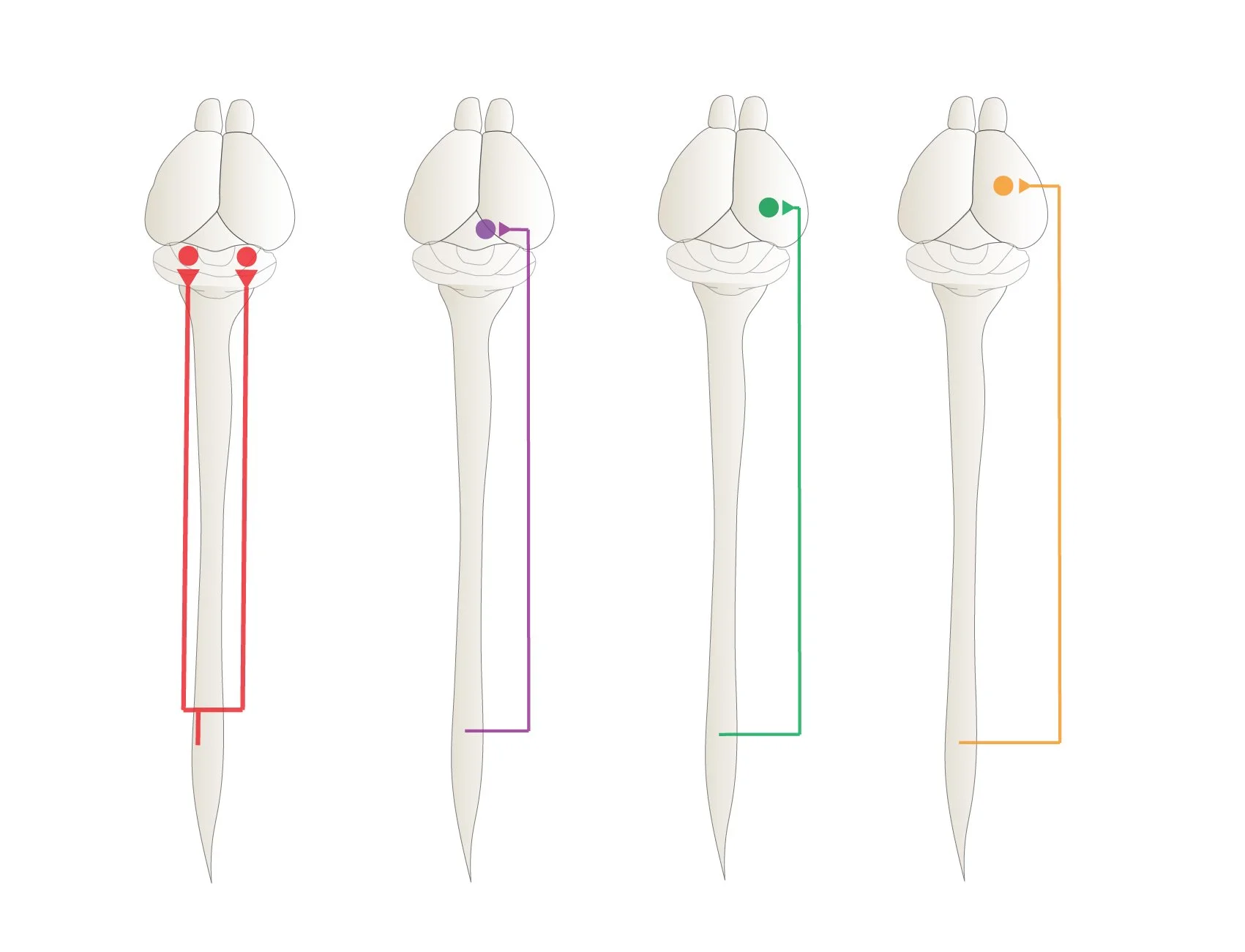
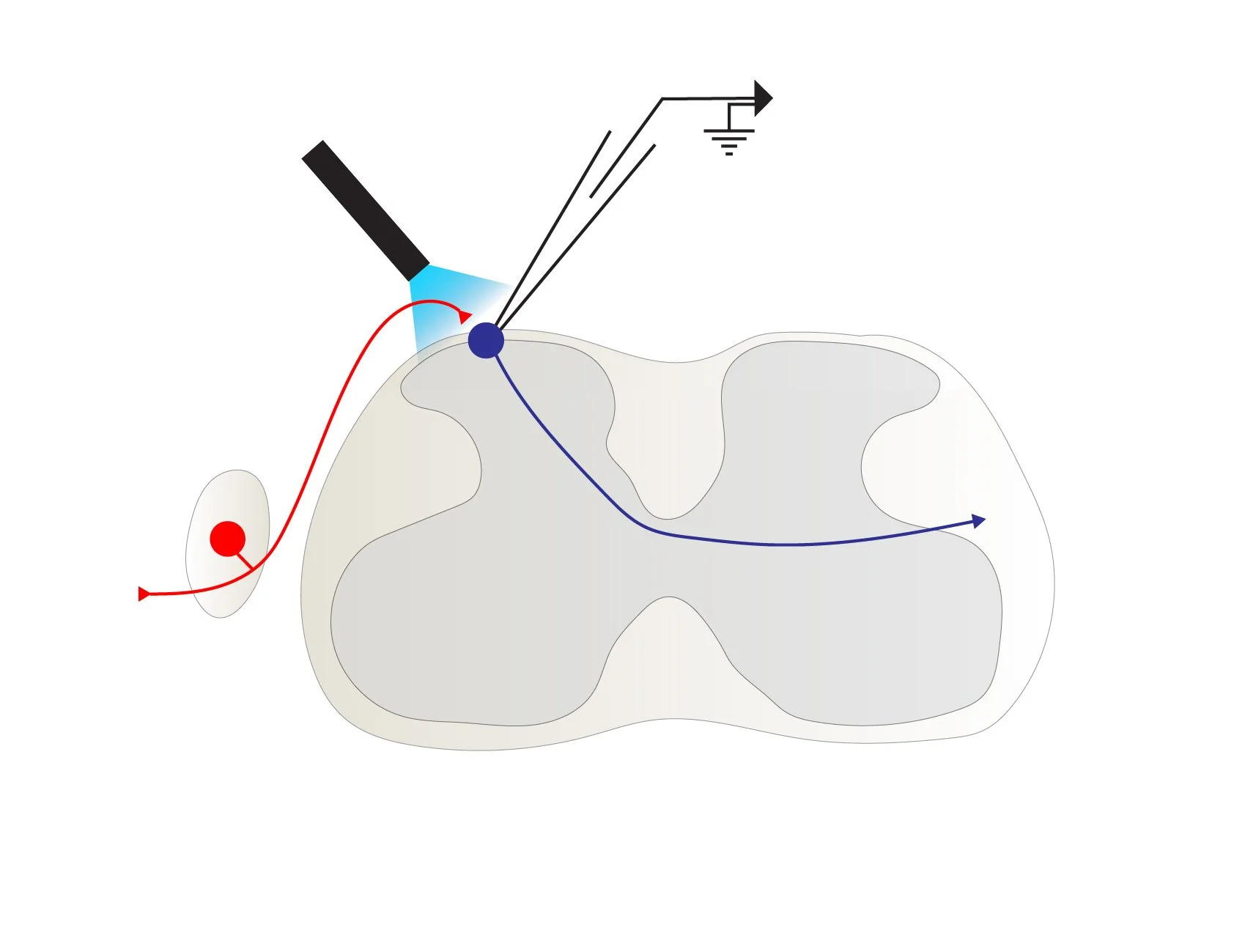
Ascending spinal pathways consist of multiple independent modules that convey touch and pain signals to many different regions in the brain, including the brainstem, pons, midbrain, and thalamus. The sophisticated structural connectivity enables precise propagation of somatosensory signals from the periphery to the brain, underlying our sensation of and reaction to touch and pain. Our lab studies how these complex ascending somatosensory circuits are wired during development and what developmental programs control their formation and organization.
Experimental approaches
-
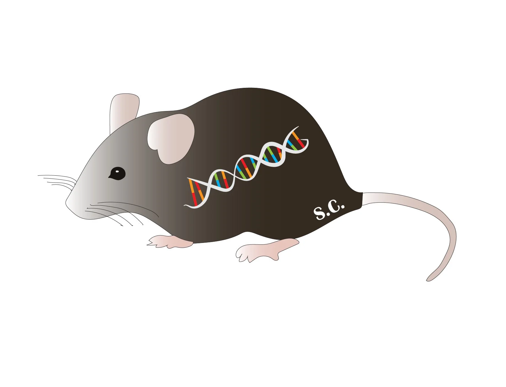
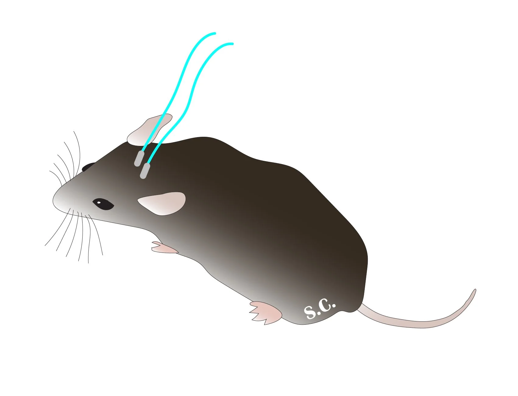
Mouse genetics
Mouse genetic tools
-
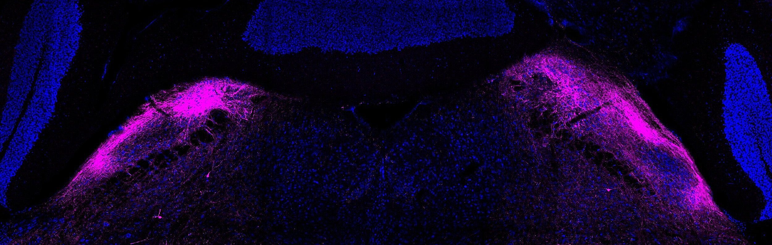
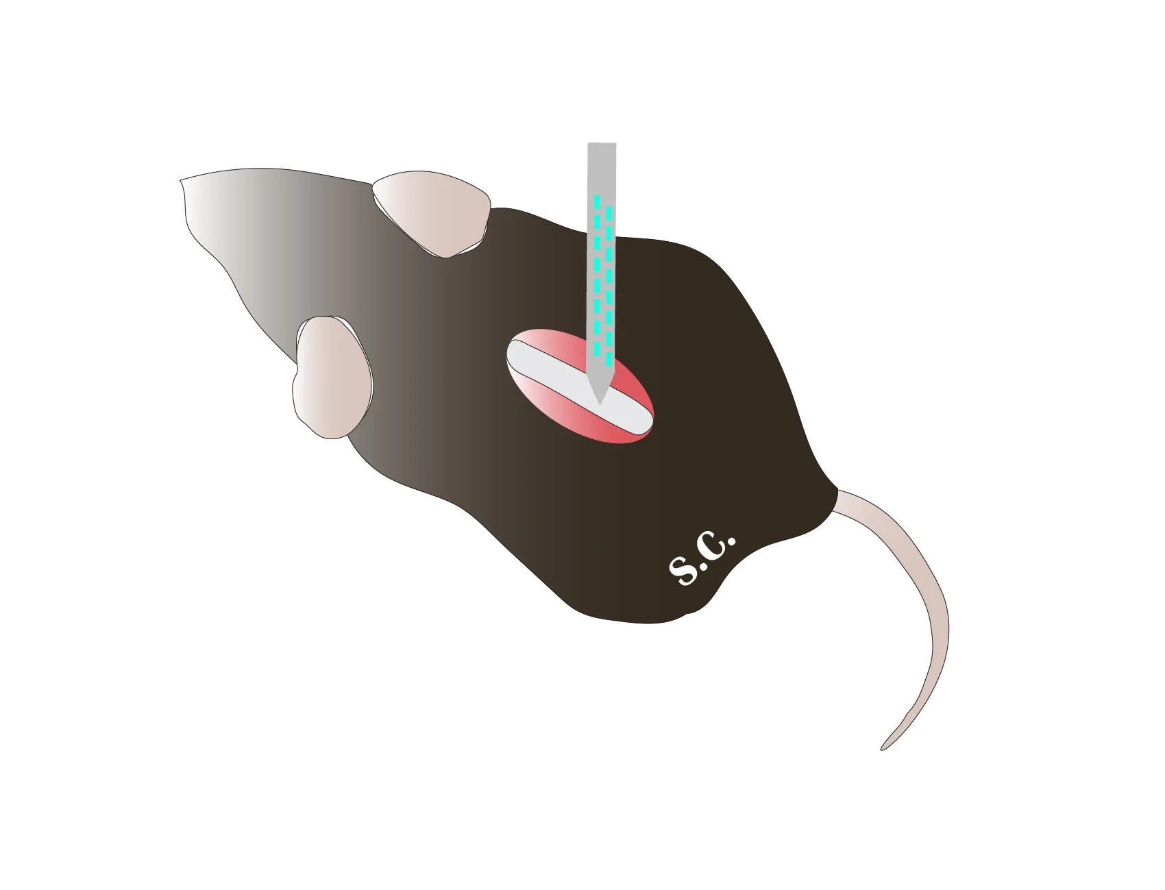
Anatomy
Intersectional strategy/Viral tracing
-
Ex vivo electrophysiology
Whole-cell patch-clamp recording
-
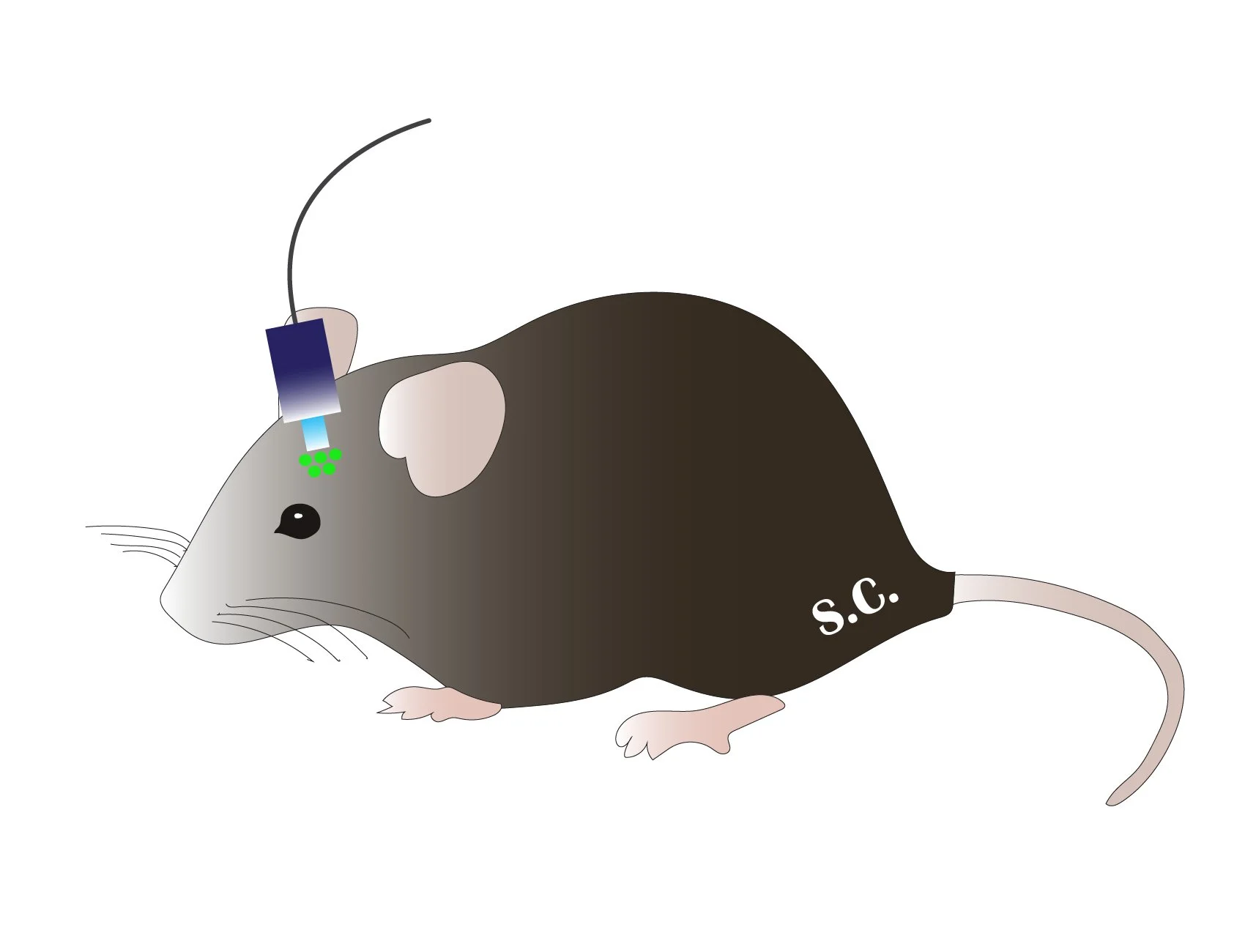
Behavior
Optogenetics/Chemogenetics
-
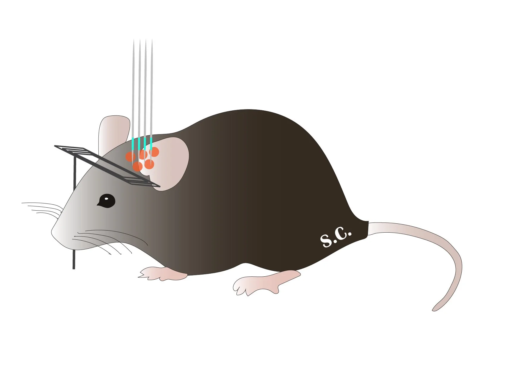
In vivo electrophysiology
Multielectrode array (MEA) recording
-
In vivo electrophysiology
Neuropixel recording
-
In vivo calcium imaging
Fiber photometry
-
In vivo calcium imaging
Miniscope imaging